Blood
Function of each part:
The Heart
The heart has 4 chambers. The upper chambers, or atria, collect the blood which then gets pumped to the ventricles through valves and then from the ventricles to the rest of the body.
The right side of the heart receives oxygen- poor blood from the rest of the body while the left side of the body receives oxygen- rich blood from the lungs and delivers that to the rest of the body.
The depolarization and contraction of the heart is controlled by the SA-node in the right atrium, also known as the pace maker.
The steps of pumping blood are as follows:
Function of each part:
- red blood cells
- transport oxygen throughout the body with the pigment hemoglobin
- white blood cells
- fight off infection with T cells and B cells and produce antibodies
- platelets
- clotting and formation of scabs
- plasma
- transportation of dissolved gasses, nutrients, and waste products.
The Heart
The heart has 4 chambers. The upper chambers, or atria, collect the blood which then gets pumped to the ventricles through valves and then from the ventricles to the rest of the body.
The right side of the heart receives oxygen- poor blood from the rest of the body while the left side of the body receives oxygen- rich blood from the lungs and delivers that to the rest of the body.
The depolarization and contraction of the heart is controlled by the SA-node in the right atrium, also known as the pace maker.
The steps of pumping blood are as follows:
- The SA-node cells generate depolarisztion, which then spreads out over the atria to the atrio-ventricular node.
- The atria then contract, pushing blood into the ventricles.
- The electrical conduction passes via the Atrio-ventricular node to the bundle of His, which divides into right and left branches and then spreads out from the base of the ventricles across the myocardium.
- This leads to a contraction of the ventricles, forcing blood up and out into the pulmonary artery (right) and aorta (left).
- The atria then re-fill as the myocardium relaxes.
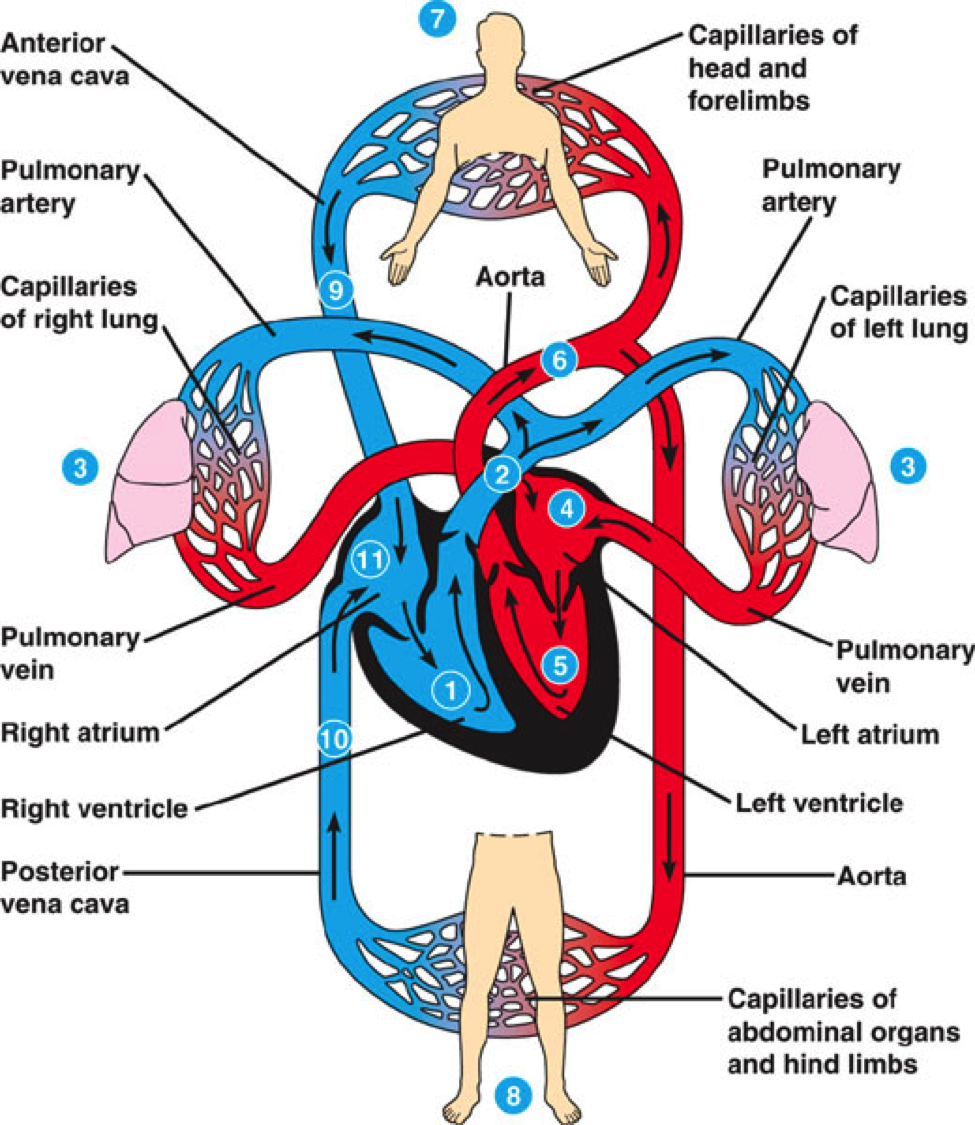
The Blood Vessels
There are 3 major types of blood vessels: arteries, capillaries, and veins. Arteries carry oxygen- rich blood throughout the body. The only artery that does not is the pulmonary artery which carries deoxygenated blood to the lungs. Capillaries are the smallest and most common blood vessels. Almost every tissue contains capillaries. Their main function exchanging gasses, nutrients, and waste products. Veins serve the opposite function of arteries- they bring deoxygenated blood to the heart from the cells of the body. Veins don't have as much pressure as arteries, so they rely on gravity and skeletal muscle contractions in order to fully pump through the body.
Blood vessels are the highway of the body. They allow for blood to travel throughout all of the body in order to maintain stable conditions. Different vessels have different wall thicknesses depending upon the purpose of the vessel (arteries are thicker, for example, than capillaries because arteries contain blood pumping at a higher pressure).
resources:
http://www.wkcardiology.com/Patient-Information/Physiology/
https://www.le.ac.uk/pa/teach/va/anatomy/case1/frmst1.html
http://www.innerbody.com/image/cardov.html
images:
http://mrborden.weebly.com/uploads/4/9/0/5/49053175/202336636.png
http://www.wkcardiology.com/Patient-Information/Physiology/
https://www.le.ac.uk/pa/teach/va/anatomy/case1/frmst1.html
http://www.innerbody.com/image/cardov.html
images:
http://mrborden.weebly.com/uploads/4/9/0/5/49053175/202336636.png
